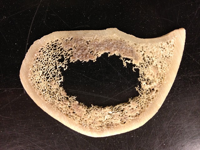
Home » Without Label » Bone Cross Section - "Bone Cross Section" for Radius Digital Science on Behance / Cross section of a human bone showing bone marrow, spongy bone and blood vessels.
Bone Cross Section - "Bone Cross Section" for Radius Digital Science on Behance / Cross section of a human bone showing bone marrow, spongy bone and blood vessels.
Bone Cross Section - "Bone Cross Section" for Radius Digital Science on Behance / Cross section of a human bone showing bone marrow, spongy bone and blood vessels.. Related posts of bone cross section labeled bones of ankle and foot on x ray. J = π*(t 4 − t 4)/32, and z = j/(t/2), where t is the external bone diameter and t is the diameter of the medullary cavity (external diameter minus total cortical thickness) at the midshaft of maximum bone length. A bone is a rigid organ that constitutes part of the vertebrate skeleton in animals. Can you identify the primary and secondary haversian systems, central canals and bone lamellae? The large dark spots are passages for blood vessels and nerves.
Vector illustration scheme of bone cross section. Scientific evidence suggests that a vegan diet might be associated with impaired bone health. This is a high power photo of a single haversian system. As the names suggest compact bone looks compact and the spongy bone looks like sponges. A hip structural analysis study osteoporos int.
David R. Nelson - Cross Section of Beef Marrow Bone from img-cache.oppcdn.com .modifications made by lies van rompaey. Diagram labeling the structure of a bone. I am not an expert on this subject, so i was wondering if anyone could put their input on this image. Browse 9,121 bone cross section stock photos and images available, or search for bone marrow or bone structure to find more great stock photos and pictures. This is a short tutorial using blender 2.8 that shows how to create a bone cross section and using images to create the textures.hope you enjoy and please su. It consists of two layers; Derivative works of this file: Image.shutterstock.com if you look at the cross section of a long bone under a microscope, the rings of bone immediately internal to the periosteum of the bone are called _____.
Related posts of bone cross section labeled bones of ankle and foot on x ray.
This photo shows a cross section through bone. (micrograph provided by the regents of university of michigan. Human back muscles and bones 12 photos of the human back muscles and bones human back muscles and bones, bone, human back muscles and bones The central tubular region of the bone, called the diaphysis, flares outward near the end to form the metaphysis, which contains a largely cancellous, or spongy, interior. On examining a cross section of any bone, it is composed of two kinds of bony tissue: This is known as the periosteum. This is a short tutorial using blender 2.8 that shows how to create a bone cross section and using images to create the textures.hope you enjoy and please su. Related posts of bone cross section labeled bones of ankle and foot on x ray. Beautiful tooth cross section illustration, deep blue background and sparkling lights around. Can you identify the primary and secondary haversian systems, central canals and bone lamellae? A hip structural analysis study osteoporos int. A bone is a rigid organ that constitutes part of the vertebrate skeleton in animals. Cross sections of bones and growth plates will result in many more knife defects.
Human back muscles and bones 12 photos of the human back muscles and bones human back muscles and bones, bone, human back muscles and bones Compact bone, spongy bone, and bone marrow. It consists of two layers; Browse 53 bone marrow cross section stock photos and images available, or search for bone cross section or bone cells to find more great stock photos and pictures. Translated into dutch.the original can be viewed here:
Bone | Cross section of long bone Leitz Orthoplan, PL FL 4 ... from live.staticflickr.com Diagram labeling the structure of a bone. Histology slide courtesy of william l. This is known as the periosteum. As the names suggest compact bone looks compact and the spongy bone looks like sponges. Image.shutterstock.com if you look at the cross section of a long bone under a microscope, the rings of bone immediately internal to the periosteum of the bone are called _____. Diagram with articular cartilage, marrow, spongy bone, medullary cavity, endosteum, diaphysis, and periosteum. This is a cross section through decalcified bone. Photomechanical print page item number:
This is a retouched picture, which means that it has been digitally altered from its original version.modifications:
Cross section of a bone : Select from premium bone cross section of the highest quality. Compact bone cross section courtesy: If i can teach you one thing about how to draw the back of a person, it's that it's absolutely crucial to understand the position of the scapula bones (shoulder in this tutorial, we will go over the bones and major muscle groups you will need to know to draw the. For example, if i missed labeling anything, or any parts of the bone are missing. Human bone, cross section diagram of femur showing osteon, veins, marrow. J = π*(t 4 − t 4)/32, and z = j/(t/2), where t is the external bone diameter and t is the diameter of the medullary cavity (external diameter minus total cortical thickness) at the midshaft of maximum bone length. There is a printable worksheet available for download here so you can take the quiz with pen and paper. This is a high power photo of a single haversian system. Human back muscles and bones 12 photos of the human back muscles and bones human back muscles and bones, bone, human back muscles and bones Cross sections of bones and growth plates will result in many more knife defects. .modifications made by lies van rompaey. On examining a cross section of any bone, it is composed of two kinds of bony tissue:
Browse 4,275 bone cross section stock photos and images available, or search for human bone cross section to find more great stock photos and pictures. This is a retouched picture, which means that it has been digitally altered from its original version.modifications: There are trabeculae in spongy bone which gives its sponge like appearance. Derivative works of this file: This is a cross section through decalcified bone.
BBC - GCSE Bitesize Science - Endoskeletons and ... from www.bbc.co.uk Photomechanical print page item number: .modifications made by lies van rompaey. As the names suggest compact bone looks compact and the spongy bone looks like sponges. Human bone, cross section diagram of femur showing osteon, veins, marrow. This is a retouched picture, which means that it has been digitally altered from its original version.modifications: Diagram with articular cartilage, marrow, spongy bone, medullary cavity, endosteum, diaphysis, and periosteum. The central tubular region of the bone, called the diaphysis, flares outward near the end to form the metaphysis, which contains a largely cancellous, or spongy, interior. Find the perfect bone cross section stock photos and editorial news pictures from getty images.
A bone is a rigid organ that constitutes part of the vertebrate skeleton in animals.
Compact bone is the outer layer and the spongy bone forms the inner layer. This is a retouched picture, which means that it has been digitally altered from its original version.modifications: Items portrayed in this file depicts. A property of the cross‐sectional area that represents the magnitude of the greatest bending rigidity of the section (cm 4). As the names suggest compact bone looks compact and the spongy bone looks like sponges. A bone is a rigid organ that constitutes part of the vertebrate skeleton in animals. Internal structure of a human long bone, with a magnified cross section of the interior. Histology slide courtesy of william l. This is a cross section through decalcified bone. Diagram labeling the structure of a bone. Smartdraw includes 1000s of professional healthcare and anatomy chart templates that you can modify and make your own. Each bone in your body is made up of three main types of bone material: Cross section of a femur bone showing the anatomical structure including cancellous bone and marrow.